The properties of 2-D transition metal dichalcogenides are attracting a great deal of interest, and one of the reasons is their catalytic activity. In particular, better catalysts are needed to exploit the potential of water electrolysis—splitting water into its component elements—to provide sustainable energy storage.
"MoS2 is one of the most promising precious rare metal-free catalysts for the hydrogen evolution reaction (HER)," point out Yasufumi Takahashi, Mingwei Chen, and Tomokazu Matsue and their colleagues at Kanazawa University and other collaborating institutions in Japan, the US and the UK in their recent Angewandte Chemie International Edition report. The work highlights the role of "scanning electrochemical cell microscopy" for engineering the catalytic properties of these 2-D materials.
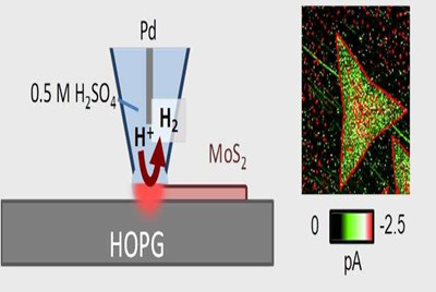
As the researchers point out, scanning electrochemical microscopy has already proved useful in investigations of the catalytic activity of MoS2 monolayers, which have focused on the effects of strain, as well as the metallic versus semiconducting properties of different microstructural phases of MoS2 on HER catalysis. These studies used a microscale electrode to probe the sample for electrochemical activity as a function of location with high spatial resolution, on account of the microscale dimensions of the electrode.
In their scanning electrochemical cell microscopy studies, Takahashi, Chen, Matsue and colleagues use a nanopipette as a local, moveable electrochemical cell to probe the electrochemical activity on the surface instead of an ultramicroelectrode. They highlight the "reproducible and reliable technique for fabricating nanoprobes together with fast electrochemical characterization due to its small capacitive current" as additional advantages of this form of the characterization technique.
The researchers used a nanopipette with a 20 nm radius to study triangular monolayers of MoS2 with a 1H microstructural phase, as well as heterostructures of MoS2 and WS2. Each flake had a side length of around 130 nm. The measurements revealed changes in catalytic activity where edges, terrace features and heterojunctions between MoS2 and WS2 were located, which agrees with the suggestions of previous reports. In addition, aging the sample had a noticeable effect, particularly at the edges.
The researchers conclude that their study demonstrates how it is possible to evaluate the local HER activity of catalytic samples using scanning electrochemical cell microscopy. They suggest that the technique can be a "powerful tool" for engineering the phase and structure of 2-D transition metal dichalcogenide samples for applications in catalysis.
